

Breast cancer is the second most common cause of cancer-related deaths in women, and the disease will affect an average of one in eight women during her lifetime. Osteoporosis is another common condition among women—they are more likely to develop the disease—and it affects more than 10 million adults, especially after age 50, in the U.S. Because we know that early detection leads to more effective treatment, we use the most advanced diagnostic techniques available to detect these diseases, and then use that information to develop a treatment plan that is right for each woman.
Our diagnostic services include the latest 3D mammography, digital mammography, ultrasound, breast biopsy and DXA bone densitometry. Our technologists, who are certified by the American Society of Radiologic Technologists and have additional certification in mammography, perform all mammograms at the UPMC Washington Women’s Center. Our fellowship-trained technicians also have extensive training specific to women’s imaging and mammography.
UPMC Washington Women’s Center
80 Landings Drive 2nd Floor, Suite 201, Washington, PA
Phone: (724) 223-3313
Screening services are also performed at the following locations:
Diagnostic Center – Peters Township
UPMC Greene
Screening Mammogram
A screening mammogram is a routine exam to check for breast cancer in women who have no signs or symptoms. The United States Preventive Services Task Force recommends that women ages 50-74 have a screening mammogram once every two years. However, before age 50, women should speak with their provider about a screening schedule that makes sense for them, taking into account family and personal history, among other factors.
After a screening mammogram is performed, a technologist views the results for technical quality. Then, a radiologist interprets the images. Your provider may recommend additional screenings or tests, if needed.
Diagnostic Mammogram
A diagnostic mammogram is an exam for those who are experiencing signs or symptoms of breast cancer. The mammogram is performed and interpreted the same day. Your provider may perform additional mammogram views and/or ultrasounds, if needed.
Diagnostic mammograms are performed when a patient has:
- a history of breast cancer
- a need for follow-up regarding a prior abnormality
- a new lump in the breast(s) or underarm(s)
- an irregularity that was discovered during a screening mammogram
- nipple discharge
- pain in the breast(s) or nipple area
- thickening or swelling in part of the breast(s)
How do I prepare for a mammogram?
Once you have scheduled your mammogram, follow these guidelines to prepare for your procedure:
- If a previous mammogram was done at a facility other than UPMC Washington, request those images be sent to the UPMC Washington Women’s Center to avoid any delay in interpretation.
- Refrain from wearing perfume, talcum powder and deodorant the day of your mammogram, as these products can interfere with the quality of the imaging.
- Bring your mammography prescription from your doctor to your appointment, as well as your health insurance card and photo ID.
What is 3-D mammography?
3D mammography, whether used during routine or diagnostic screening, allows doctors to examine breast tissue one layer at a time. It uses high-powered computing to convert digital breast imaging into a stack of very thin layers, or slices, building what is essentially a 3D mammogram.
What is the difference between 3-D mammography and conventional digital mammography?
Conventional digital mammography is still one of the most advanced technologies available for the diagnosis of breast cancer. However, it shows all the breast tissue at a glance, where one feature can hide in the shadow of another. 3D mammography eliminates or reduces the superimposing shadow, and a radiologist can search through the breast in 1-millimeter layers, almost like turning pages in a book. This makes it easier to determine if there’s any cause for concern and decrease the chance for a tumor to hide behind overlapping tissue. 3D mammography does not eliminate being called back because of irregular results; however, it greatly decreases the chances of being called back for additional imaging, because all of the breast tissue can be seen more clearly.
Will my insurance pay for a 3-D mammogram?
Your insurance may not cover 3D mammography rather than traditional mammography. Please call your individual insurance company prior to your appointment to verify your individual coverage plan.
Who should get a 3-D mammogram?
3D mammograms are approved for all women who can undergo a screening or diagnostic mammogram. It is helpful for all women, regardless of breast density.
What happens during a 3D mammogram?
Currently, 3D mammography complements, but does not replace, the standard 2D digital mammogram. It only takes a few more seconds for each view. Both mammograms are done at the same time, using the same compression and mammography system. The X-ray mammography machine sweeps over the breast from one side to the other in a slight arch motion, and a series of digital images are obtained and processed to generate 3D images.
Are there any risks with 3D mammography?
Just as with conventional mammography, 3D mammography uses very low doses of radiation. The total radiation dose is similar to a conventional digital mammogram and is safely below the guidelines approved by the American College of Radiology.
Do I need a physician referral for a 3-D mammogram?
You will need a referral/prescription from your physician to schedule your digital screening or diagnostic mammogram, but your doctor does not have to specifically order the 3D mammogram. If you are interested in having a 3D mammogram, please notify the scheduler when you call to make an appointment. The appointment must be scheduled as a 3D mammogram to provide appropriate resources.
How do I prepare for a mammogram?
Once you have scheduled your mammogram, follow these guidelines to prepare for the exam:
- Try not to schedule your mammogram appointment for the week before you get your period or during your period to avoid having tender or swollen breasts.
- If a previous mammogram was done at a facility other than WHS, request those images be sent to the WHS Women’s Center to avoid any delay in interpretation.
- Refrain from wearing perfume, talcum powder and deodorant the day of your mammogram, as these products can interfere with the quality of the imaging. Plan to wear a two-piece outfit the day of your mammogram (e.g., no dresses, jumpers), so you do not have to get completely undressed during the exam.
- Bring your mammography prescription from your doctor to your appointment, as well as your health insurance card and photo ID.
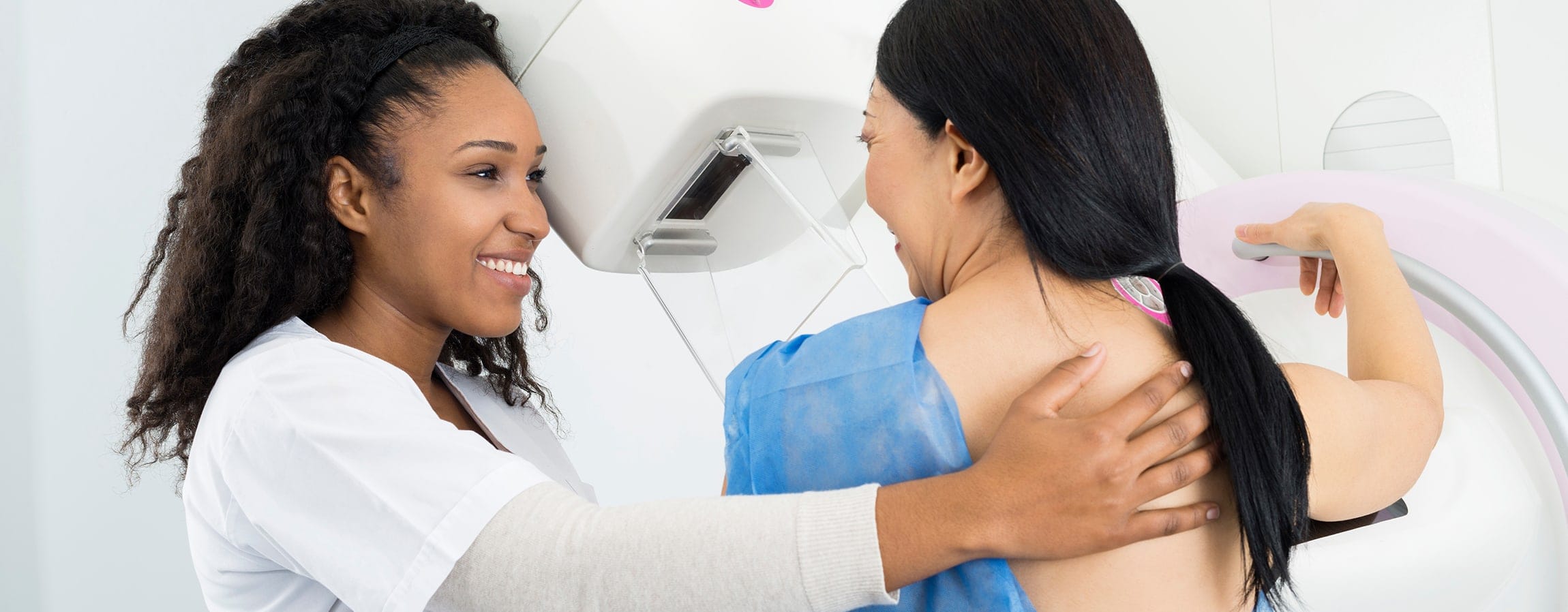
What Will Happen During the Mammogram?
- Your breast will be positioned in the mammography unit and placed on a platform for compression.
- Your breast will be gradually compressed so that all breast tissue can be seen and examined, and small abnormalities are not obscured.
- It is important to remain still and hold your breath during the mammogram, so that the image is not blurred.
- The compression of your breast may be uncomfortable, especially for women who have very sensitive breasts, but compression aids in diagnosis.
- Allow up to 2 hours for your entire diagnostic mammogram appointment.
How will I receive my test results after my mammogram?
The UPMC Washington Women’s Center staff of fellowship-trained and board-certified radiologists will review and interpret your mammography images. Once your mammogram is completed, you may resume your normal activities unless your physician directs otherwise. If you have any additional questions, please contact the UPMC Washington Women’s Center at (724) 223-3313.
You and your physician will be notified of the results of your exam. If the radiologist has identified an area of concern, WHS will contact you directly and ask that you return for additional testing, if needed.
Ultrasound imaging, or sonography, uses high-frequency sound waves to produce real-time pictures of the body. Similar to an ultrasound exam that is used for expectant mothers, physicians can use ultrasound imaging to produce images of the breast by using a transducer probe to produce an image of the breast tissue on a computer screen.
Ultrasound allows radiologists to take a closer look into breast tissue, to determine whether an abnormality is:
- a fluid-filled benign cyst
- concerning or cancerous
It is important to remember that finding an abnormality in the breast does not necessarily mean it is cancerous. Breast ultrasound can be used if mammography or MRI are not conclusive enough for your physician to determine the cause.
You may have a breast lump or some other change in your breast. Most breast lumps or other changes are not cancer. However, to be sure, you will be scheduled for a nonsurgical breast biopsy.
A nonsurgical breast biopsy describes various techniques that do not require surgery to obtain samples of cells or tissue to establish a precise diagnosis. This is the way to determine if the abnormality is benign (not cancerous) or malignant (cancerous).
There are a variety of nonsurgical biopsy techniques available today. A nonsurgical breast biopsy requires advanced equipment to precisely locate and remove a small sample of tissue. A radiologist and technologist specially trained in this technique will perform the procedure. Once they obtain the tissue sample, a pathologist (a doctor specially trained to review tissue samples) examines it. The tissue sample must be prepared and processed before a final diagnosis can be made. This usually takes approximately one week.
In general, these biopsies are performed with the use of a local anesthetic, and patients have a quick return to normal activities.
How do I prepare for my breast biopsy?
IMPORTANT:
- If you take aspirin or blood thinners on an everyday basis, you must stop taking them for 5 days before your exam
- In addition, if you take blood thinners, you will need to arrive 1 1/2 hours before your exam to have blood work done in the Meadows Landing Outpatient Center Lab, which is within our suite. You must have a prescription from your doctor for this lab work. Please take the doctor’s slip with you to the lab.
- If you feel you will need more than local anesthesia, please contact your doctor for additional pain or calming medication prior to your scheduled procedure.
- Please eat normally on the day of your test.
- We recommend that you bring a tight-fitting or athletic support bra to wear after the procedure to minimize movement of the breast and discomfort.
Is a breast biopsy painful?
Everyone has a slightly different pain threshold. A local anesthetic will be used to numb your breast to minimize the discomfort you might feel.
Breast MRI (Magnetic Resonance Imaging) is an advanced imaging tool that, when used in conjunction with annual screening mammograms, gives your physician the best chance of detecting cancer.
Breast MRI can also be useful for determining the best course of treatment for women who have been newly diagnosed with breast cancer. Breast MRI does not take the place of mammography, but is used to supplement the information that mammography provides.
Breast MRI may be ordered to evaluate:
- abnormal breast tissue identified through mammography or ultrasound
- a mass that can be felt during a manual breast examination
- breast implants
- very dense breast tissue
A breast MRI biopsy may also be required to make a definitive diagnosis. MRI-guided breast biopsy is a minimally invasive procedure, requiring a shorter recovery time than traditional surgical biopsy.
How do I prepare for my breast MRI?
When you schedule your appointment, you will be asked a series of screening questions to determine if there is anything in your medical history that might prevent you from having an MRI.
If you have a history of renal impairment, diabetes or hypertension, you will need to obtain a blood test (GFR) to determine if your kidneys are functioning well enough to receive gadolinium, an intravenous contrast agent typically used in breast MRI. Please have your blood testing done at least 48 hours prior to your MRI appointment.
If your mammogram or breast ultrasound was done at a facility other than the UPMC Washington Women’s Center, it is very important that you bring your films or a CD of the exams with you to your appointment or arrange to have them sent over.
What should I know about my breast MRI?
The day of the examination:
- You may eat and drink normally.
- You may take your medications.
- Please leave valuables and jewelry at home.
- Bring any prior mammography or ultrasound films with you if they were not performed at the UPMC Washington Women’s Center.
- Report directly to the Admitting Department on the 2nd floor (main lobby).
- You will be directed to the MRI department on the ground floor of the UPMC Washington Hospital.
- Plan on being in the department for up to two hours.
- You will read and sign a consent form.
- You will change into a special wraparound patient gown.
- Remove any metal objects, including your watch, body piercings, glasses and hearing aids. You will be assigned a locker to store your personal items.
What will happen during a breast MRI?
- You will be asked to lie on your stomach on a table that slides into the MRI scanner.
- Your breasts will hang through specially-cushioned openings.
- During your scan, the technologist will monitor you through a large window and communicate with you via an intercom.
- All MRI scanners generate some vibrations and make some knocking and pinging noises while they are acquiring the images.
- Ear plugs or headphones for noise reduction are available for your use.
- You must hold still while the MRI is being performed, because any movement will cause the images to be out of focus and the test may need to be repeated.
Is it safe to have an MRI?
It is not safe for you to have an MRI if you have any of the following:
- breast tissue expander
- cochlear implant
- implanted defibrillators
- metallic fragments in your eyes
- pacemaker
- surgical clips from surgeries in the prior six weeks
Some things might or might not be compatible with an MRI. Our medical staff will determine if it is safe for you to have an MRI if you have any of the following:
- aneurysm clips
- medication patches
- metallic implants
- neurostimulators
- nonremovable body piercings
- pregnancy
- prosthetic heart valves
- recent surgery or procedure
- spinal cord stimulator
- stents
- surgical staples or clips
- tattoos
Breast cancer can spread through the lymph ducts and lymph nodes to other areas of the body. To determine if the cancer has spread, and to determine how aggressive treatment needs to be, surgeons remove the lymph nodes under the arm. Surgeons use a less-invasive method, called sentinel node biopsy, to determine if breast cancer has spread beyond the breast. Sentinel node biopsy causes fewer complications and side effects than traditional axillary lymph node dissection, where a surgeon:
- makes an incision underneath the arm and removes the bulk of the lymph node tissue that drains from the breast
- on average, removes 10 to 15 lymph nodes
- sends samples to the laboratory, where a pathologist looks at the lymph nodes to determine if any of them contain cancer
With removal of a high number of lymph nodes, there is a greater chance of:
- arm swelling
- decreased mobility
- pain, numbness and nerve injury
Furthermore, axillary node dissection usually requires an overnight stay at the hospital, whereas sentinel node biopsy does not.
Through sentinel node biopsy, the surgeon only removes one to three lymph nodes. The sentinel lymph node is the first node that filters fluid from the breast, and experts believe that malignant cells reach the sentinel node first. If the sentinel is free of cancer cells, it is unlikely that the other nodes are positive.
Sentinel node biopsy can also lead to a more accurate assessment of whether cancer has spread. In a traditional axillary dissection, the pathologist makes fewer cuts in each of the lymph nodes to look for cancer. When the pathologist receives only the sentinel node, he or she makes many cuts through the node to look for cancer. This also gives them the capability to use more highly specific stains.
- If the sentinel node is negative, there is a greater than 95% chance that the other lymph nodes are also cancer free. If the results are positive, the surgeon may then perform an axillary node dissection to see how many other nodes are affected.
- With increased screening, we are finding smaller cancers sooner, which means less chance of cancer spreading to the lymph nodes. Therefore, a higher number of axillary dissections are going to be negative. The sentinel node biopsy eliminates unnecessary surgery and risks.
- Sentinel node biopsy is covered by Medicare and most private insurances.
One of the most effective ways to detect osteoporosis is through a bone density exam (DXA scan). This is performed with state-of-the-art technology and is painless and noninvasive.
While the patient lies still on a padded table, the technologist moves the arm of the equipment to the area of the body to be examined. A computer, which is connected to the equipment, displays an image of the examined area. The images are analyzed to obtain the bone density measurements. The results are then interpreted by the radiologist.
As always, our emphasis at UPMC Washington Hospital is on quality care, and our caring and professional staff is very sensitive to the patient’s needs. The X-ray exposure is minimal, and the procedure takes approximately 30 minutes. During the procedure, our technologist collects information about your current lifestyle and physical condition. A full report of the test results is sent to your ordering physician.
The exam determines:
- the calcium mineral content of the examined areas by comparing each patient’s results with others within the same age and gender in a national database
- the risk for developing bone fractures of the hip and spine
- the rate of bone loss because of aging and menopause
- the therapeutic effect of medications commonly prescribed for treating osteoporosis
Physician referral is needed for a DXA scan. Please call (724) 223-3313 for an appointment. Patients who are not sure about the terms of their insurance should call and ask if dual energy X-ray absorptiometry (DXA) is covered.
Women’s Center Support Services
Breast Patient Navigator
Patient navigation is a process by which an individual—a patient navigator—guides patients through and around barriers in the complex cancer care system, to help ensure timely diagnosis and treatment.
UPMC Washington Women’s Center provides certified breast patient navigation throughout the patient journey. The certified breast patient navigator guides patients through diagnosis and treatment by answering questions, providing information about available services and amenities, and ensuring a comfortable experience for the patient.
Additional information: ‘Navigator’ Assists Breast Cancer Patients
Breast Cancer Support Group
The UPMC Washington Women’s Center hosts a Breast Cancer Support Group the second Tuesday of every other month. The meetings are for current and previous breast cancer patients. There are various topics for discussion and speakers throughout the year.
Meetings are held from 6 p.m. to 7:30 p.m. at the Wilfred R. Cameron Wellness Center. For more information, call (724) 223-3313.
To find a provider who can help you navigate all aspects of women’s health, use our physician finder or call (724) 250-4310.